
Figure 2.
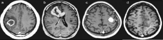
Four Different Patients with GBM that Illustrate the Heterogeneity in the Anatomic Lesion. The contrast-enhanced axial T1-weighted (TR, 600 msec; TE, 14 msec) images demonstrate variegated appearance of GBM: (a) rim-enhancing mass with central necrosis in the right parietal lobe with surrounding edema; (b) irregularly enhancing mass that crosses the corpus callosum; (c) well-circumscribed homogeneously enhancing mass in the left frontal lobe with no associated edema; (d) ill-defined infiltrative mass in the left medial frontal lobe with no appreciable necrosis. (Adapted from: Nelson and Cha, 2003).