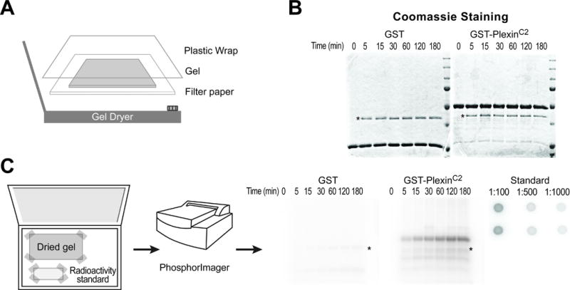
Fig. 2. Detection of phosphorylated protein using a phosphorimager screen.

(A) To dry a destained gel, put the gel on a wet filter paper, wrap with a plastic film and place on the center of a heated gel dryer connected to a vacuum generator.
(B) A representative dried gel shows two major protein bands, a substrate with the strongest intensity and a protein kinase with a weaker intensity (asterisk, PKA in this example).
(C) Left: place a dried gel and a radioactivity standard spotted on filter paper in a phosphorimager cassette and tape the corners. Middle: detect radioactivity using a phosphorimager scanner. Right: representative images of an autoradiograph from the dried gel and a radioactivity standard. GST-PlexinC2 protein, but not GST alone, is phosphorylated. Note that the signal intensity increases over the reaction time. Autophosphorylated PKA is also detected (asterisk). The numbers in the radioactivity standard indicate dilution factors.
