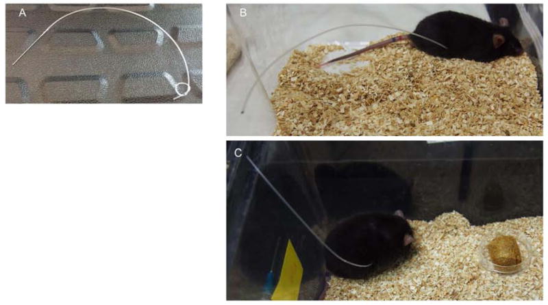
Figure 1. Dialysis catheter and placement in mice.
A) Representative image of the custom peritoneal dialysis catheter is shown. PD catheters consisted of 8 to 10 inches of PE50 sterile tubing with a 2 centimeter loop, and 6 to 8 holes. B) and C) Images of catheter placement in mice. Mice were singly housed and the PD catheter was tunneled through the dorsum of the mouse to allow for easy access for PD fluid exchanges and to be out of reach so that the mouse would be unable to chew or pull out the catheter.

