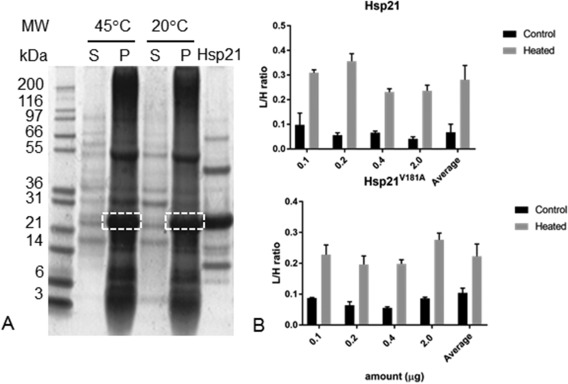
Figure 2.

Quantification of Hsp21 protein associated with thylakoid membranes at increased temperature. A. Outline of the experimental set‐up in which thylakoid membranes were incubated at 20°C and 45°C with unlabeled (14N) Hsp21, and samples spiked with known amounts of 15N‐labeled Hsp21 before sample processing. By centrifugation pellet (P) with thylakoid membranes and supernatant (S) were separated, followed by SDS‐PAGE gfractionation. Samples excised (dashed rectangles) were subjected to LC‐MSMS to determine the L/H‐ratios. B. Triplicate samples of thylakoid membranes were processed as described in panel A and the relative amount of Hsp21 in thylakoid membranes incubated with Hsp21 at 20°C (Control) and 45°C (Heated) determined as L/H‐ratio. Results are shown for Hsp21 protein (upper panel) and for Hsp21V181A, a non‐dodecameric mutational variant (lower panel).
