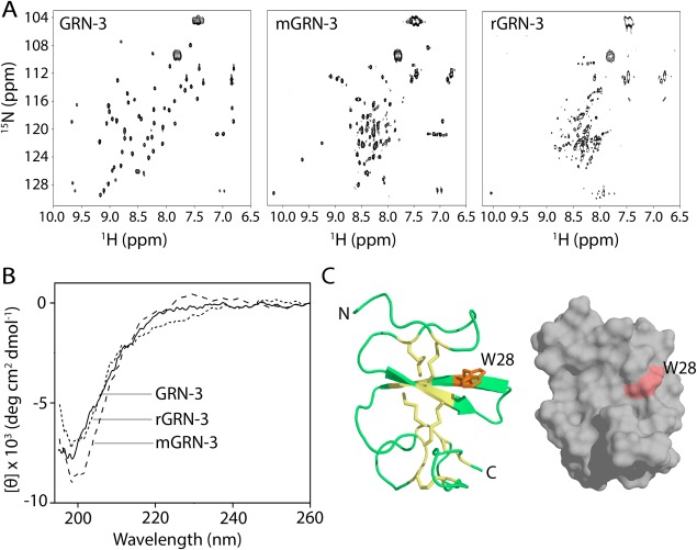
Figure 4.

Structural characterization of GRN‐3, mGRN‐3, and rGRN‐3. (Α) 1H‐15N HMQC NMR spectra GRN‐3, mGRN‐3, and rGRN‐3. (B) Far‐UV circular dichroism (CD) spectra of GRN‐3 (solid line), mGRN‐3 (dashed line), and rGRN‐3 (short dashed line). Both GRN‐3 and rGRN‐3 show minima at 198 nm typical of random coil conformation whereas mGRN‐3 shows a minimum at 202 nm and a maximum at ∼230 nm corresponding to a PP‐II helix. (C) Structural model (ribbon and space‐filled representations) of GRN‐3 obtained using I‐TASSER with disulfide bonds represented as sticks (yellow) and W28 (orange) exposed on the surface.
