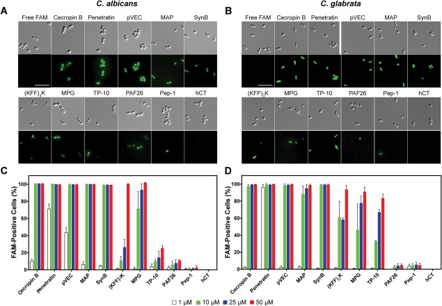
Figure 1.

Translocation of FAM‐labeled CPPs into Candida cells. DIC and FAM fluorescence images for translocation into (A) C. albicans and (B) C. glabrata. Flow cytometry data for translocation into (C) C. albicans and (D) C. glabrata. Cells were incubated for 1 h at 30°C with 25 µM peptides for imaging or serial dilutions of peptides (1–50 µM) for flow cytometry. Surface‐bound peptides were removed by trypsin prior to imaging or flow cytometry, and controls with free FAM were included. For (A) and (B), scale bar = 10 µm. For (C) and (D), error bars represent the standard error of the mean for three separate experiments (N = 3).
