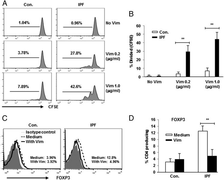
FIGURE 2.
Supplementation of PBMC cultures with native vimentin induced proliferation of CD4 T cells from IPF patients. (A) Representative line graphs showing CFSE dilution in gated CD4 T cells from controls (Con.) and IPF patients. Dose dependence was evident in increasing vimentin concentrations from top to bottom. (B) Quantitation of aggregate CFSE assays in Con. (n = 10) and IPF (n = 15). Representative line graphs showing downregulation of CD4 T cell FOXP3 (C) and aggregate compilations of the same (D). Cultures in these studies were stimulated or not with vimentin (1 μg/ml). **p < 0.01.

