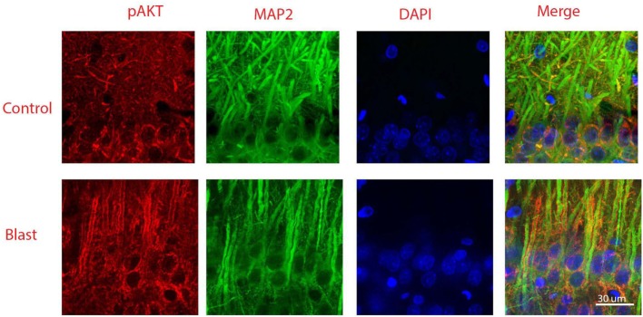
Figure 5.
Co-immunostaining of pAkt S473 and microtubule-associated protein 2 (MAP-2) in the CA1 region of ipsilateral hippocampus 1 week after primary blast (25 psi) exposure. Phosphorylated Akt was shown in red and MAP-2 was shown in green. Phosphorylated Akt is mainly stained along dendritic membranes. DAPI was used to counterstain nuclei (blue). Magnification: 400×.

