Fig. 1.
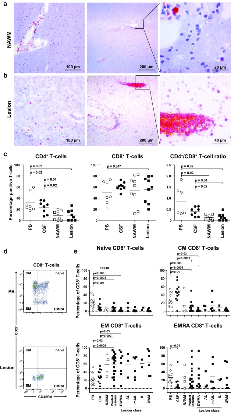
CD8+ T cells in normal-appearing and diseased white matter tissues of MS patients preferentially express an effector memory phenotype. a, b Parenchymal T cells are selectively detected in active MS lesions. 10-µm cryostat sections of paired (a) normal-appearing white matter (NAWM) and (b) white matter lesion (WML) of two representative patients of five MS patients analyzed. CD3 expressing cells (T cells) were stained with 3-amino-9-ethylcarbazole (red color) and counterstained with hematoxylin (blue color). Whereas perivascular T cells were detected in both NAWM and WML, parenchymal T cells were exclusively detected in active WML of MS patients. c Percentages of CD4+ and CD8+ T cells, and CD4+/CD8+ T-cell ratio, are shown for paired PB (open circles), CSF (closed circles) and histologically defined as NAWM (open squares) and WML (filled squares). Lymphocytes were isolated from paired peripheral blood (PB), cerebrospinal fluid (CSF), NAWM and WML (lesion) from patients with advanced MS (n = 17) and subjected to multiplex flow cytometry. Gating procedure of CD4+ and CD8+ T cells is shown in Online Resource 3. d CD8+ T cells were subdivided in naïve (CD27+CD45RA+), central memory (CM; CD27+CD45RA−), effector memory (EM; CD27−CD45RA−) and terminally differentiated effector memory (EMRA; CD27−CD45RA+) T cells. Gating procedure is shown for representative paired PB and WML-derived CD8+ T cells. e The frequency of naïve, CM, EM and EMRA CD8+ T cells is shown for paired PB, CSF and white matter brain tissues that were immunohistologically classified as NAWM, diffuse white matter abnormalities (DWMA), active lesions (AL), mixed active/inactive lesions (mIAL), inactive lesion (IL) or unconfirmed white matter tissues (UWM) (see Online Resource 3 for criteria applied for MS WM classification). Horizontal lines represent the mean frequencies. Wilcoxon matched pairs test was used to calculate significance

