Fig. 2.
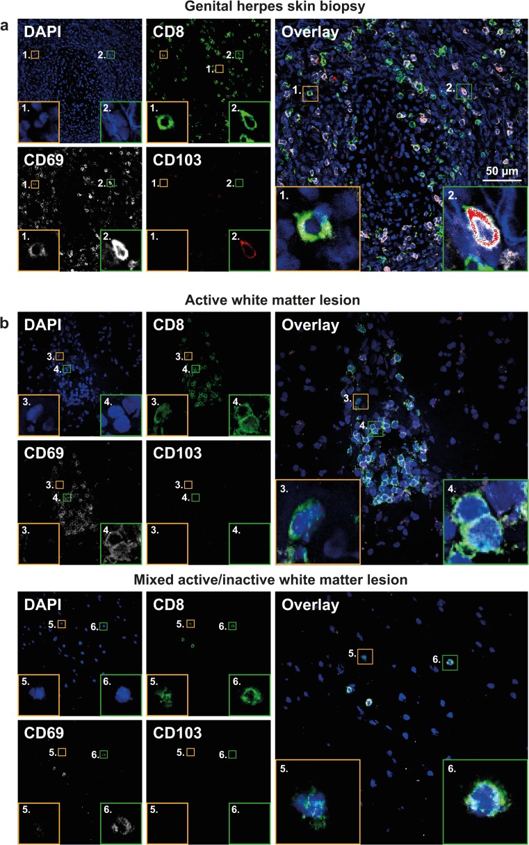
CD8+ T cells in white matter lesions of MS patients preferentially express CD69, but not CD103. a, b Representative triple immunofluorescent stainings on 8-μm cryosections of a skin biopsies from six genital herpes patients and b immunohistochemically classified white matter lesions (WML) of six MS patients. Tissues were stained for CD8 (green color), CD69 (white color) and CD103 (red color) using specific monoclonal antibodies (mAbs) and isotype specific fluorochrome-conjugated secondary antibodies. Sections were counterstained with 4′,6-diamidino-2-phenylindole (DAPI; blue color) and analyzed using a Zeiss LSM-700 confocal laser microscopy and ZEN software. Skin biopsies of genital herpes patients were stained as positive control to validate staining strategy by confirming localization tissue-resident CD8+ T cells based on differential CD69 and CD103 staining: CD8+CD69+CD103− (a; inset 1) and CD8+CD69+CD103+ T cells (a; inset 2). b In WML of MS patients, perivascular (top panels) and parenchymal CD8+ T cells (bottom panels) were incidentally CD69−CD103− T cells (b; insets 3 and 5) and predominantly CD69+CD103− T cells (b; insets 4 and 6). Size bar is indicated in top-right image

