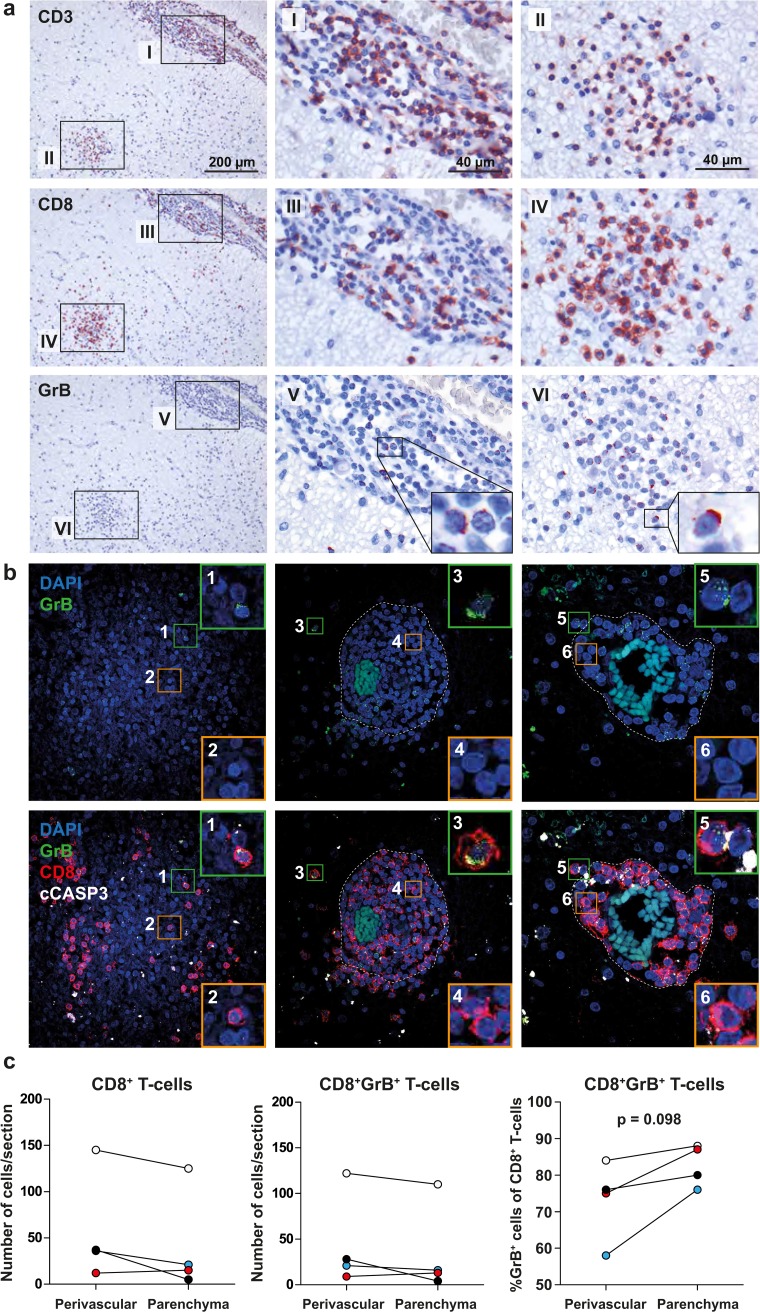
Fig. 4.
CD8+ T cells in white matter lesions of MS patients express granzyme B. a Representative stainings on 6-µm sections of a formalin-fixed and paraffin-embedded (FFPE) mixed active/inactive white matter lesion (mAIL) of one of four MS patients analyzed. CD3 (top panel), CD8 (middle panel) and granzyme B (grB) expressing cells (bottom panel) were stained with 3-amino-9-ethylcarbazole (red color) and counterstained with hematoxylin (blue color). Abundant punctate expression of grB was detected in perivascular (insets I, III and V) and parenchymal (insets II, IV and VI) CD8+ T cells. Granzyme B polarization was observed in both perivascular and parenchymal CD8+ T cells (insets V and VI). b Representative maximum intensity projections of z-stack laser confocal microscopy images of immunofluorescent triple stainings for grB (green color), CD8 (red color) and the early apoptotic cell marker “cleaved caspase-3” (cCASP3; white color). Stained sections were counterstained with DAPI (blue color). Representative stainings of three mAIL are shown of 12 immunohistochemically classified WML tissues of 10 MS patients analyzed. Punctated (inset 1) and polarized grB expression by CD8+ T cells (insets 3 and 5), as well as grB-negative CD8+ T cells are shown (insets 2, 4 and 6). Co-localization of grB and cCASP3 is observed in a cell adjacent to a CD8+ T-cell with polarized grB suggesting grB-mediated killing of the respective target cell (inset 5). Dotted line represents the glia limitans separating the perivascular space and parenchyma. c The grB-expressing CD8+ T cells were counted in the perivascular space and parenchyma of mAIL of four MS patients analyzed. Wilcoxon matched pairs test was used to calculate significance

