Fig. 5.
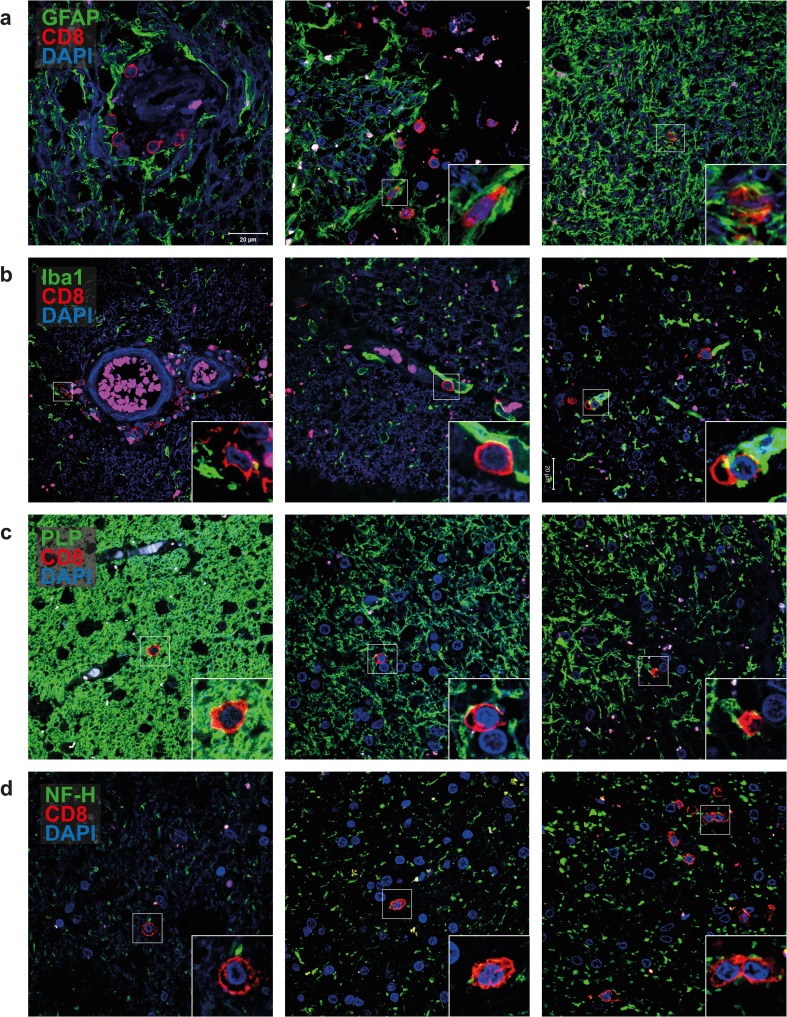
CD8+ T cells interact with all major brain-resident cell types in white matter lesions of MS patients. Representative double-immunofluorescence stainings on 8-μm sections of 12 formalin-fixed paraffin-embedded white matter lesion (WML) tissues from 10 MS patients are shown for a glial fibrillary acidic protein (GFAP: marker for astrocytes); b ionized calcium-binding adapter molecule 1 (Iba1: marker for microglia); c proteolipid protein (PLP: marker for oligodendrocytes) and d neurofilament heavy chain (NF-H: marker for neurons; all green color) combined with CD8 (red color), counterstained with 4′,6-diamidino-2-phenylindole (DAPI; blue color) and finally analyzed using a Zeiss LSM-700 confocal laser microscopy and ZEN software. Perivascular CD8+ T cells interact with astrocytes (a left and middle panel) and microglia (b left and middle panel). Parenchymal CD8+ T cells also interact with astrocytes (a right panel), microglia (b right panel) and oligodendrocytes (c) in fully myelinated (left panel) and partially demyelinated areas (middle and right panels). Parenchymal CD8+ T cells also interact with neurons (d) in areas without (left panel), with moderate (middle panel) and with prominent axonal swelling (right panel). The latter is indicative of axonal damage. Insets show specific interactions between CD8+ T cells and the major brain-resident cells analyzed for. Scale bar is indicated (a, top left panel)

