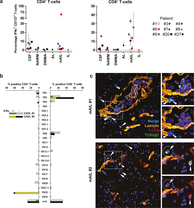
Fig. 7.
White matter lesion-derived CD8+ T cells recognize autologous Epstein–Barr virus-transformed B cells and localize in the parenchyma to form immune synapses. a Short-term T-cell lines (TCL) were generated by non-specific stimulation of T-cell recovered from paired cerebrospinal fluid (CSF) and white matter brain tissues from nine MS patients, which were immunohistologically classified defined as normal-appearing white matter (NAWM), diffuse white matter abnormalities (DWMA), active lesions (AL), mixed active/inactive lesions (mAIL) and inactive lesions (IL) (see Online Resource 3 for criteria applied for MS WM classification). The TCL were incubated with autologous Epstein–Barr virus-transformed B cell lines (autoBLCL). Next, the phenotype and frequency of autoBLCL-specific T cells was determined by co-expression of intracellular interferon gamma (IFNγ) and CD137 using multiplex flow cytometry. The frequency of autoBLCL-reactive T cells is shown as the percentage of IFNγ+CD137+ CD4+ (left panel) and CD8+ T cells (right panel). Symbols represent individual donors (n = 9; specified in the legend) and vertical lines represent the mean and standard deviation of at least two independent experiments per TCL. Significance of variation in autoBLCL T-cell reactivity was determined by ANOVA for CD4+ and CD8+ T cells separately. b TCL generated from two anatomically distinct mAIL of MS patient #6 (see Online Resource 1) were cultured with autoBLCL to assay the T-cell receptor variable β chain (TCRVβ) usage of the reactive T cells, determined by intracellular interferon gamma (IFNγ) expression, using multiplex flow cytometry. The frequency of CD4+ (left x-axis) and CD8+ T cells (right x-axis) of specific TCRVβ families (y-axis) are depicted (gray bars lesion #1, black bars lesion #2). The frequency of autoBLCL-reactive CD4+ T cells and CD8+ T cells of each TCRVβ family is shown (stacked green bars IFNγ+ T cells). Results shown are representative for two independent experiments. “Und.” refers to T cells expressing a TCRVβ chain not covered by the TCRVβ-family-specific monoclonal antibody panel used. c Triple immunofluorescence staining for TCRVβ2 (green color), laminin (orange color) and CD8 (red color) in surplus WML tissue sections (8 µm) containing WML #1 and #2, from which the corresponding TCLs shown in panel “A” were generated. Nuclei were stained with DAPI (blue color). TCRVβ2+ CD8+ T cells reside in the perivascular cuff (open arrowhead) and the parenchyma (closed arrowhead) of both distinct WML of the same patient. The majority of parenchymal T cells show polarization of both CD8 and TCRVβ2 (encircled cells in top-right insets). Images of representative stainings are shown

