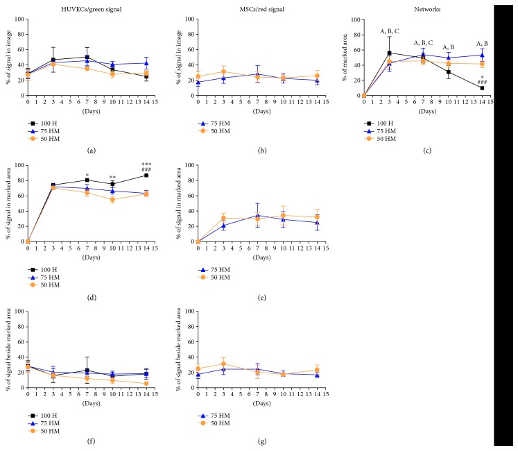
Figure 5.
Analysis of cellular networks. Image analysis was performed of mono- and cocultures (100% HUVECs (100 H), 75% HUVECs–25% MSCs (75 HM), and 50% HUVECs–50% MSCs (50 HM)) in PRP (seeding density: 2.5 × 10³ cells/μl gel) (n = 3). Networks were measured by manually marking cellular networks. The relative percentage of prestained green (HUVECs) and red (MSCs) cells was investigated for day 0, day 3, day 7, day 10, and day 14 in the following regions: relative percentage of green or red signal in the entire image (a, b), relative percentage of marked network area (c), relative percentage of green or red signal in marked network area (d, e), and the relative percentage of green or red signal beside marked network area (f, g). For 100 H monocultures, networks decreased after one week whereas in both cocultures, networks were present and stable after two weeks (c). Cellular networks were mainly made out of HUVECs (d), with a minor contribution of MSCs (e). Two-way ANOVA with Tukey's post hoc test for multiple comparison was applied to test for significant differences over time (compared to day 0: (A) 50 HM p < 0.01 (all time points); (B) 75 HM p < 0.01 (day 3) and p < 0.001 (day 7–14); and (C) 100 H p < 0.001 (day 3) and p < 0.01 (day 7)) and between different culture conditions (∗p < 0.05, ∗∗p < 0.01, ∗∗∗p < 0.001 compared to 50 HM, ###p < 0.001 compared to 75 HM). n = 3.

