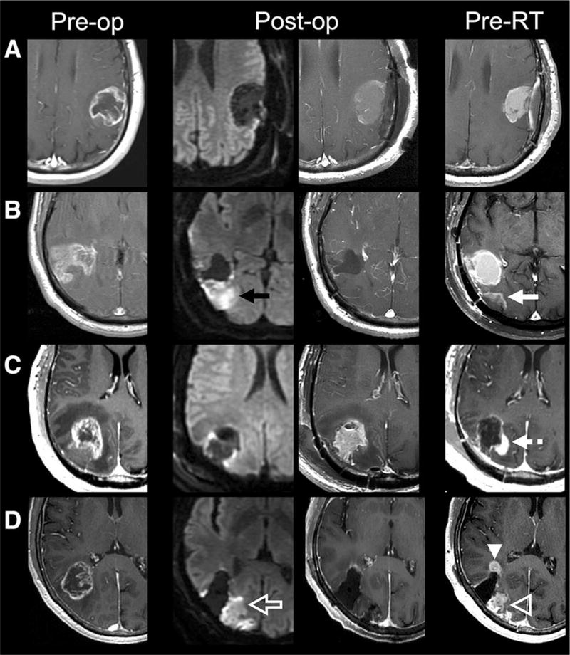
Fig. 1.
Preoperative (T1 postcontrast, first column), immediate postoperative (DWI, second column; T1 postcontrast, third column), and preradiotherapy (T1 postcontrast) axial MR images of selected patients. a Gross total resection of enhancing tumor without immediate postoperative reduced diffusion and without new contrast enhancement to suggest tumor growth at preradiotherapy MRI. b Gross total resection of enhancing tumor with reduced diffusion along the posterior margin of the resection cavity (black arrow) that demonstrates contrast enhancement on preradiotherapy MRI (white arrow) consistent with evolving postoperative injury. c Gross total resection of enhancing tumor with minimal pericavity reduced diffusion that demonstrates new nodular contrast enhancement along the posteromedial resection cavity (dashed white arrow), suggestive of tumor progression. d Gross total resection of enhancing tumor with reduced diffusion along the posterior resection cavity (open white arrow) that demonstrates contrast enhancement on preradiotherapy MRI consistent with evolving postoperative injury (open white arrowhead) as well as new nodular contrast enhancement not associated with diffusion abnormality (white arrowhead) suggestive of tumor progression

