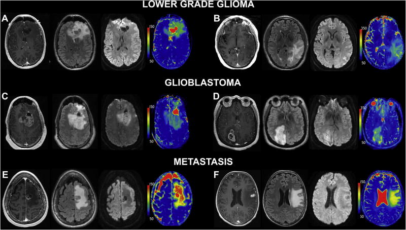
Fig. 1.
T1ρ differentiates glial neoplasm from intraparenchymal metastatic disease. Six representative patients from the three studied tumor groups; imaging from right to left: post-contrast 3D T1 SPGR, FLAIR, DWI (T2 trace image), and T1ρ color map. LGG: (A) 41-year-old man with anaplastic astrocytoma (WHO grade III) and (B) 57-year-old man with oligoastrocytoma (WHO grade II). GBM: (C) 52-year-old man and (D) 76-year-old woman with GBM (WHO grade IV). Metastatic disease: (E) 64-year-old woman with small cell lung carcinoma metastasis and (F) 67-year-old woman with metastatic breast cancer. Similar morphologic appearance is demonstrated on post-contrast 3D T1 SPGR, FLAIR, and DWI images. Markedly increased T1ρ values are observed within the NCE peritumoral edema portion of the intracranial metastasis. Conversely, the gliomas demonstrate decreased T1ρ within the NCE pertiumoral edema when compared to metastatic disease. This observation across the imaging cohort was found to be statistically significant. Note elevated T1ρ values within the increased bulk water of the cystic/necrotic components in (A), (C), and (E).

