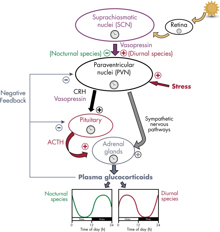
Figure 7.
Schematic representation of the control of the circadian rhythmicity of GC release in mammals. Most components of the HPA axis contain circadian oscillators. The circadian secretion of GC is dependent on the rhythmic release of ACTH and a gating process by the adrenal clock. GC secretion is also modulated by nervous signals coming from the PVN of the hypothalamus via sympathetic nervous pathways. ACTH release is controlled by the rhythmic release of CRH and vasopressin from the PVN. Rhythmic activity of the HPA axis is under the control of the master clock in the SCN, reset by ambient light via the retina. The peak of vasopressin release from the SCN to the PVN region occurs during daytime in both nocturnal and diurnal rodents. In nocturnal rats, vasopressin exerts an inhibitory action on the PVN (probably via activation of γ-aminobutyric acid-containing interneurons), thus reducing GC secretion during daytime (green curve). By contrast, in diurnal grass rats, vasopressin stimulates PVN activity (probably via activation of glutamatergic interneurons), thus increasing GC secretion during daytime (red curve). Clock symbols represent self-sustained oscillators.

