Figure 5.
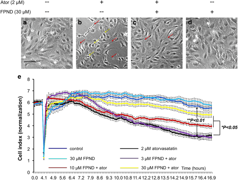
FPND significantly prevented atorvastatin-induced cell retraction and rupture of cell–cell junctions on HUVECs. HUVECs were treated with 0.1% DMSO (a and b) or 30 μM FPND (a and b) for 2 h, followed by washout and incubation with 0.1% DMSO (a and d) or 2 μM atorvastatin (b and c) for 12 h. Imaging was done with a phase contrast microscope. Yellow arrows show retracted cells; red arrows show formation of pseudopodia. Scale bar=100 μm (black color). (e) The representative cell index showed that FPND inhibited atorvastatin-induced EC contraction and rupture of cell–cell junctions. HUVECs were cultured on the E-Plate in complete medium for 48 h and pretreated with 3, 10, 30 μM FPND for 2 h, followed by washout and incubation with 2 μM atorvastatin for 24 h. ‘Ator’ in figure indicates 2 μM atorvastatin. Data presented in the bar graphs are the mean±S.D. of three independent experiments. *P<0.05 and **P<0.01 (versus the atorvastatin-alone group) were considered significantly different.

