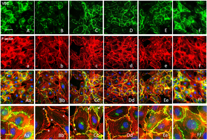
Figure 6.
FPND prevented atorvastatin-induced VEC junction dissociation and loss of VEC from cell borders. HUVECs monolayers were treated with 0.1% DMSO (A, a, Aa, A`; B, b, Bb and B`), and 5 (C, c, Cc and Cc`), 10 (D, d, Dd, Dd`), and 20 (E, e, Ee, Ee`; F, f, Ff and Ff`) μM FPND for 2 h, followed by washout and treatment with 0.1% DMSO (A, a, Aa, A`; F, f, Ff and Ff`) or 2 μM atorvastatin (B, b, Bb and B`; C, c, Cc and Cc`; D, d, Dd and Dd`; E, e, Ee and Ee`) for 12 h; 0.1% DMSO treatment for 12 h was the vehicle control (A, a, Aa, Aa`). Treatment with FPND alone (F, f, Ff and Ff`) resulted in a slight decrease in stress fiber formation but with no effect on VEC distribution or cell–cell junctions. The VEC signal was labeled with VEC-specific antibody in green color (A–F). F-actin was labeled with tetramethyl rhodamine isothiocyanate (TRITC)-phalloidin in red color and nuclei were labeled with the nuclear-specific dye Hochest 33342 in blue color (A–F). White asterisks indicate formation of scrambled knots, membrane ruffle and focal adhesion complexes. White arrows show a drastic loss of VEC from cell borders and formation of a net-like structure. White and yellow scale bars=50 and 10 μm, respectively.

