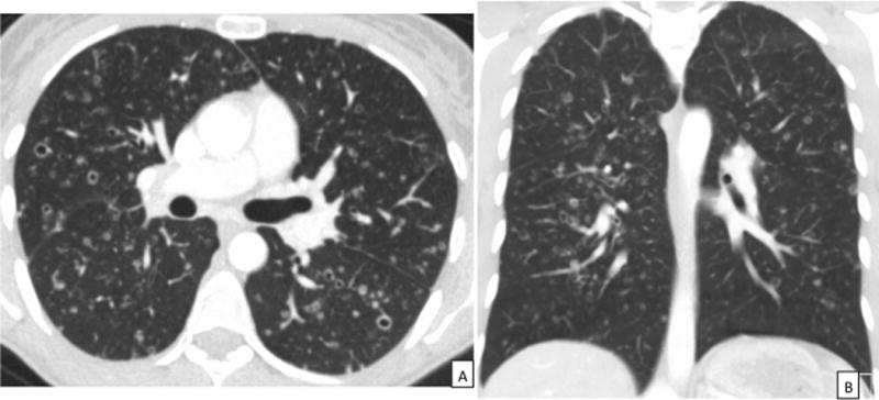
Figure 2.

CT images in a patient with PLCH. 2A: Axial CT chest demonstrating the characteristic thin-walled cysts, thick-walled cavities, and nodules in a patient with PLCH. 2B: Coronal view of the CT scan in the same patient highlighting the upper lobe predominance of radiographic abnormalities with sparing of the costophrenic sulci characteristic of PLCH. CT = Computed Tomography, PLCH = pulmonary Langerhans cell histiocytosis.
