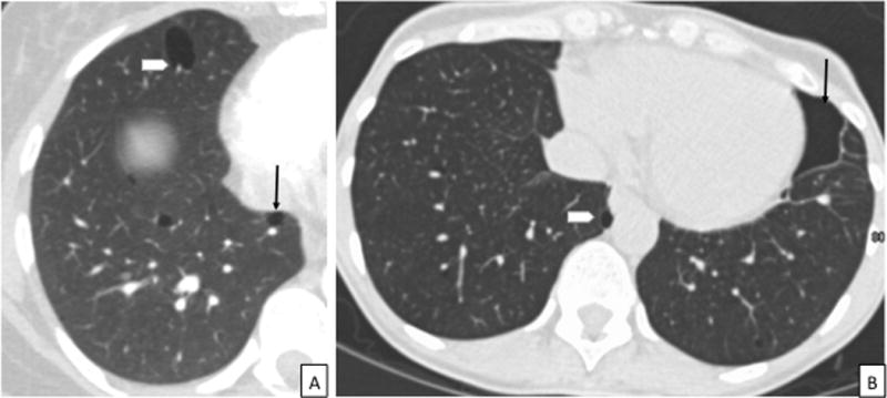
Figure 3.

CT images in a patient with BHD. 3A: Axial CT chest showing the characteristic thin-walled, lentiform cysts abutting the pleura (arrow) and pulmonary vasculature (arrowhead), in a lower lobe distribution. 3B: CT chest showing a chronic loculated left sided pneumothorax in a patient with BHD (arrow). Notice the presence of characteristic lentiform-shaped BHD cyst abutting the mediastinal pleura (arrowhead) in addition to the loculated pneumothorax. CT = Computed Tomography, BHD = Birt-Hogg-Dubé syndrome.
