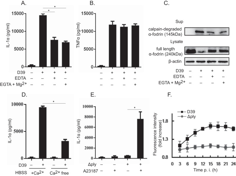
FIG 5.
Involvement of elevated intracellular calcium concentrations in IL-1α secretion. (A and B) Macrophages were infected with D39 and/or the Δply strain at an MOI of 1. Subsequently, cells were treated with 10 mM EDTA and 10 mM EGTA plus 0.7 mM Mg2+ 3 h after infection in the presence of gentamicin, and the culture was continued for a further 21 h. The culture supernatants were collected, and the levels of IL-1α (A) and TNF-α (B) were measured. (C) The full-length form and the proteolytic fragment of α-fodrin were detected by Western blotting in the cell lysate and the culture supernatant. β-Actin was utilized as a loading control. (D) Alternatively, macrophages were infected with D39 for 6 h and cultured in calcium-free (Ca2+-free) or calcium-containing (+Ca2+) RPMI 1640 medium. HBSS, Hanks' balanced salt solution. (E) Similarly, 10 μM the reagent A23187 was added 3 h after infection in the presence of gentamicin, and the culture was continued for a further 21 h. The culture supernatants were collected, and the level of IL-1α was measured by an ELISA. Data represent the means and the standard deviations of results from triplicate assays and are representative of data from three independent experiments. (F) Macrophages were infected with the D39 and Δply strains at an MOI of 1. The intracellular calcium concentration was monitored every 3 h after infection for 24 h by using a Fluo-4 NW calcium assay kit (Invitrogen) according to the manufacturer's instructions. All of the experiments were repeated three times. Statistical significance was determined by Student's t test. *, P < 0.05.

