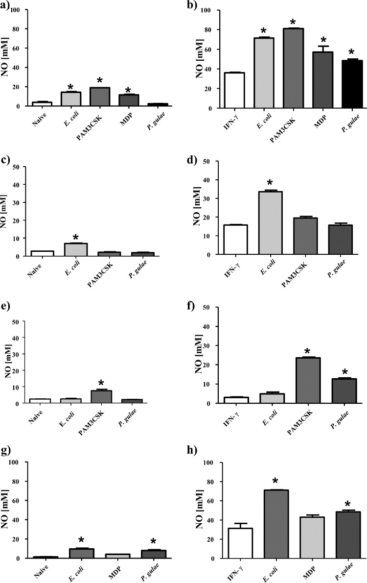
FIG 3.
Nitric oxide production by macrophages in response to Porphyromonas gulae whole cells. WT iMACs, TLR2−/− iMACs, and TLR4−/− iMACs (105 cells) were incubated overnight with or without IFN-γ to prime the M1 macrophage phenotype or to give unprimed M0-Mϕ, respectively. The unprimed and M1-primed macrophages were then incubated with viable P. gulae (MOI of 180:1) for 2 h and washed with DMEM containing antibiotics. E. coli LPS and Pam3CSK4 were used as TLR4 and TLR2 control ligands, respectively. The negative control was macrophages that were not incubated with TLR ligands or P. gulae cells. After 24 h, the level of nitric oxide expression was measured using the Griess reaction. The assay was repeated twice on each of three biological replicates (n = 6). (a) WT Mϕ; (b) WT M1-Mϕ; (c) TLR2−/− Mϕ; (d) TLR2−/− M1-Mϕ; (e) TLR4−/− Mϕ; (f) TLR4−/− M1-Mϕ; (g) NOD2−/− Mϕ; (h) NOD−/− M1-Mϕ. *, P < 0.05 compared to the corresponding unstimulated control.

