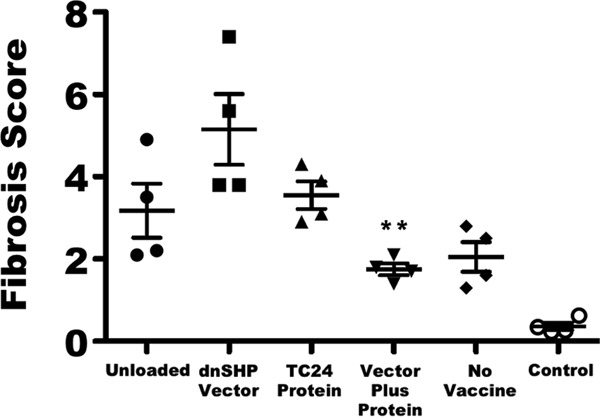
FIG 6.

Quantitation of Masson's trichrome staining. To quantify cardiac fibrosis, 5-μm sections of heart tissue were stained with Masson's trichrome stain. Images of three to five representative sections from each mouse were captured at a ×100 magnification by using a Fisher Micromaster microscope and Micron software. Images were evaluated by a reviewer who was blind to the treatment groups and analyzed by using ImageJ FIJI software to quantify the area of fibrosis and the total tissue area. The level of fibrosis in the vector-plus-protein group was significantly lower than those in all other infected groups as determined by one-way ANOVA (**, P < 0.01). Data for uninfected age-matched control mice are shown at the far right for comparison. Error bars indicate SEM.
