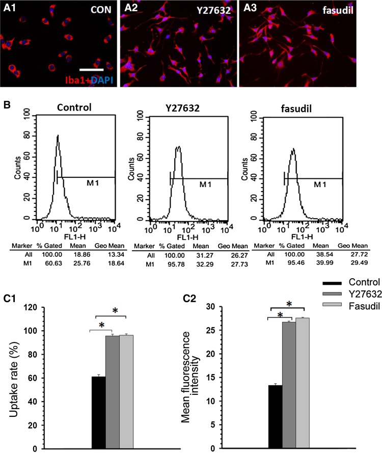
Fig. 2.
Y27632 and fasudil changed cell morphology and enhanced uptake activity in BV2 microglia. (A) Double staining with Iba1 (red) and DAPI (blue) 1 h after treatment in the control (A1), Y27632 (A2), and fasudil (A3) groups (scale bar, 25 μm). (B) Representative examples of flow cytometric analysis of control-, Y27632-, and fasudil-treated BV2 cells at 1 h. (C) The data indicated that Y27632 and fasudil stimulated uptake activity in BV2 cells. The percentage of BV2 cells that took up FITC-dextran (C1) and the mean fluorescence intensity of FITC-dextran (C2) in BV2 cells increased after Y2762 and fasudil treatment. The results are expressed as the mean ± SEM (n = 5, *P < 0.01, Y27632 vs control group, fasudil vs control group).

