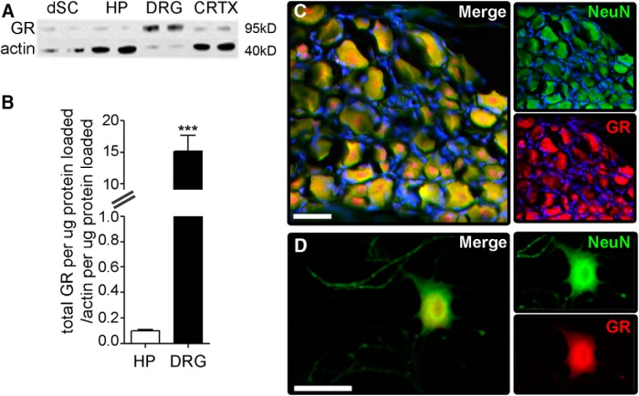
Figure 1.
DRG neurons express high levels of GR. A, GR expression was analyzed via Western blotting using protein isolated from homogenates of dorsal spinal cord (dSC), hippocampus (HP), DRG, or cerebral cortex (CRTX). B, GR expression in DRG was >15-fold higher than in hippocampus (p < 0.0001); N = 4 per group, mean and SEM are shown. C, Immunohistochemical staining for GR (red) in sections of whole DRG or (D) purified adult mouse DRG neurons in vitro reveal that GR is localized to neurons. NeuN is in green and DAPI in blue. Scale bar, 40 μm.

