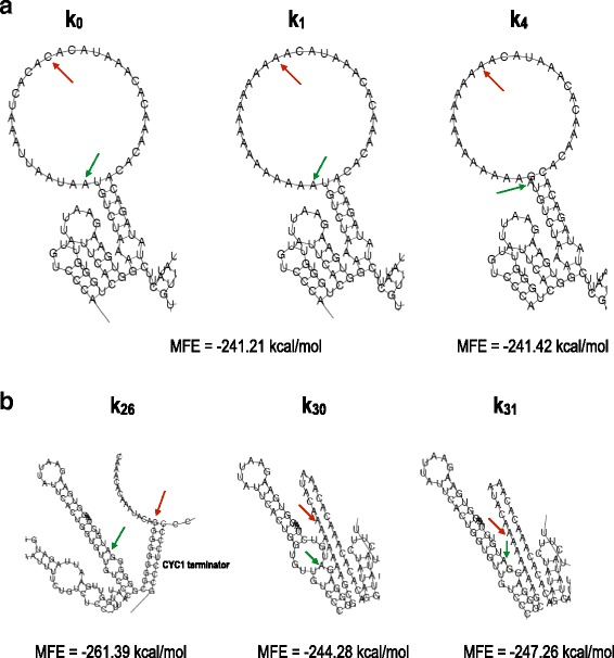
Fig. 4.

mRNA secondary structures. a A giant hairpin is present in the mRNA secondary structure corresponding to the MFE of both k 0 and k 1. The hairpin loop contains the −15…−1 region. The portion of the 5′-UTR in our analysis is free from any pairing interactions in its wild-type configuration (k 0) and in that theoretically optimized for high protein expression (k 1). The loop of the giant hairpin is reduced in k 4 owing to the base-pairing interaction between the guanine at position −1 and the cytosine at position −31. In every mRNA structure presented, a green arrow indicates position +1, and a red arrow indicates position −15. b The disruption of the giant hairpin induces a decrease in the MFE of the mRNA secondary structure. k 26 and k 31 are associated with the lowest MFEs computed in our analysis. The two sequences contain multiple guanines in the extended Kozak sequence involved in pairing interactions with the CDS. A similar pattern is also present in k 30. Here, however, a second mini-loop around the START codon provokes an increase in MFE. The MFE of k 26 is substantially lower than those of k 30 and k 31 because of the presence of another stem due to pairing interactions between the upstream region and the CYC1 terminator. Nevertheless, the fluorescence levels of k 30 and k 31 are only approximately 1.2-fold higher than that of k 26
