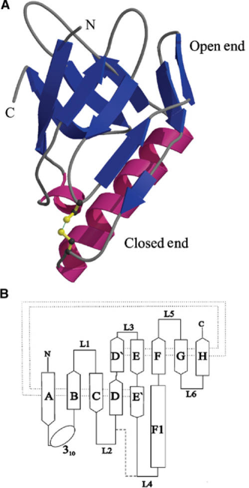
Figure 1.

Structure of MxiM. (A) A ribbon representation of the protein with the β-structure shown in blue and the helical domains shown in magenta. (B) The fold topology of MxiM is shown where the β-strands of the antiparallel β-sheet are shown as arrows and labeled A–H. The α-helix is shown as a rectangle and labeled F1. The dotted line represents the hydrogen bonding interactions between the β-strands and the dashed line represents the single disulfide bond in the structure.
