
Figure 6.
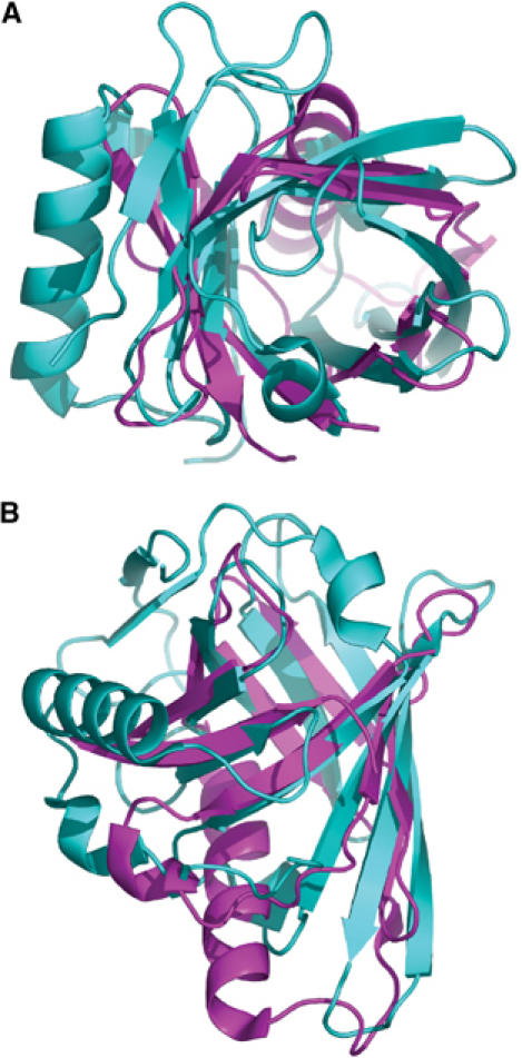
Superposition of MxiM (magenta) and bacterial lipocalin Blc (cyan; PDB code: 1QWD) carried out using PyMol (DeLano, 2004). The C-terminal helix of Blc, which is positioned at the side of the β-barrel, was excluded from the figure for clarity. (A) View looking down the internal cavities. (B) Side view rotated 90° from view (A). Blc had the highest structural similarity to MxiM based on a DALI search (Holm and Sander, 1995) where the root mean square deviation (r.m.s.d.) was calculated to be 3.1 Å (86 common Cα atoms). While there is β-strand overlap between the two structures for all eight strands of Blc, the proposed insertion of the α-helix in MxiM divides two of its β-strands resulting in two additional β-strands.