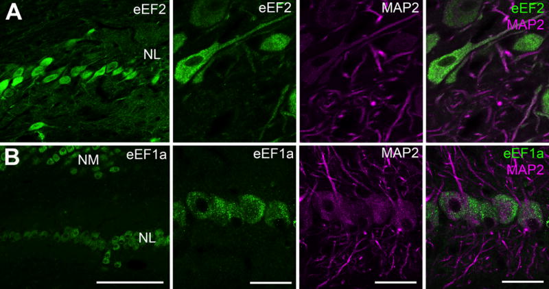
Figure 6.

Subcellular distribution of elongation factors 1a and 2 in NL examined by immunocytochemistry. A: Double labeling of phosphorylated eEF2 (p-eEF2) and MAP2, showing the distribution of eEF2 in both cell bodies and dendrites of NL neurons. Dendritic level of p-eEF2 varies between branches. B: Double labeling of eEF1a and MAP2. Strong immunoreactivity for eEF1a was observed in NL cell bodies, while no detectable staining was found in NL neuropil regions containing dendrites. Abbreviations: NM, nucleus magnocellularis; NL, nucleus laminaris; MAP2, microtubule-associated protein 2; eEF1a, eukaryotic elongation factor 1a; eEF2, eukaryotic elongation factor 2. Scale bar = 100 μm (left column) and 20 μm (all other columns).
