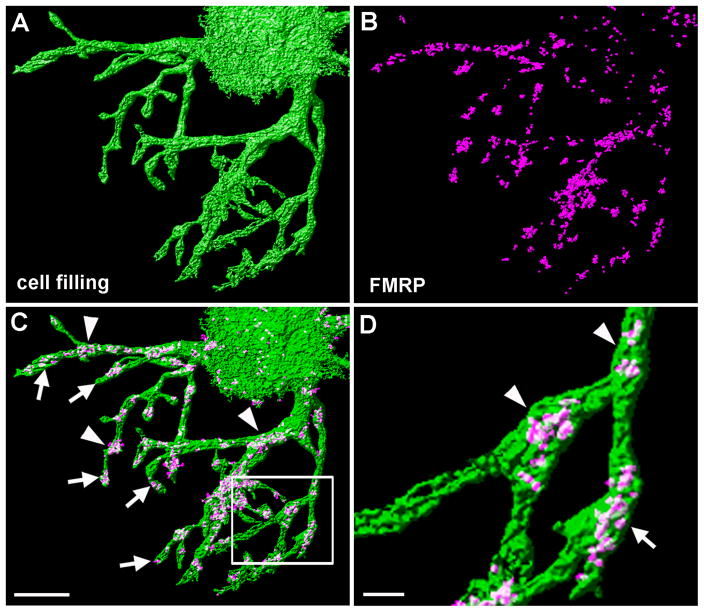
Figure 11.
Dendritic localization of FMRP in individual NL neurons. A, Surface rendering of a single NL neuron that was filled with a fluorescent dye. The image here shows the ventral dendritic arborization and a part of the soma (right up corner). B, Overlapped FMRP immunoreactivity on dye-filled dendritic branches. Non-overlapping immunoreactivity was removed using the Object Colocalization Analysis function of the Huygens software for visualization. C, Merged image of A and B showing strong accumulation of FMRP immunoreactivity in branch points (arrowheads) and enlarged distal tips (arrows). D, Closer view of an isolated dendritic branch in the box in C. Scale bar = 10 μm in C (applies to A–C) and 2 μm in D.

