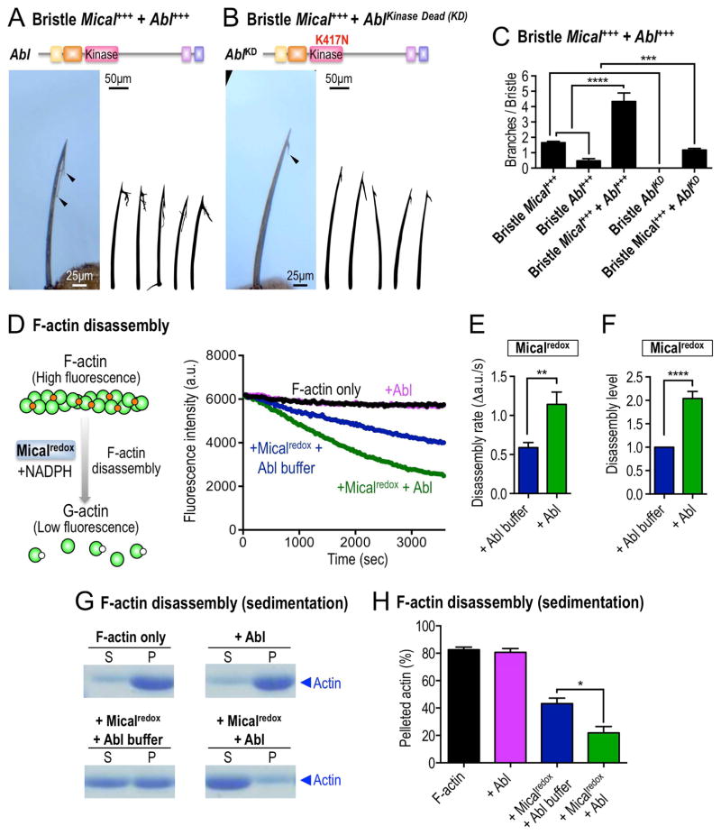
Figure 2. Abl augments Mical-mediated actin disassembly.
(A–C) Abl modulates Mical-mediated actin alterations and cellular remodeling in vivo in a kinase-dependent manner. (A, C) Increasing the bristle levels of Abl enhances Mical-induced F-actin/cellular remodeling and generates bristles with longer and more complex branches (e.g., arrowhead, compare with Figure 1F). (B–C) Bristle specific expression of a kinase inactive mutant of Abl (AblKD) suppresses Mical-induced F-actin alterations/cellular remodeling (e.g., arrowhead; compare with Figure 1F). n≥33 bristles/genotype; mean±s.e.m; t-test; ****P≤0.0001; ***P≤0.001. (D–F) Abl uses its kinase activity to directly promote Mical-mediated F-actin disassembly. Pyrene-labeled actin depolymerization assays using purified proteins, in which Mical’s ability to disassemble F-actin could be followed in real-time, reveal that F-actin disassembles (decreasing fluorescence) in the presence of Micalredox (blue, preincubated with Abl kinase buffer). Pre-incubation of Micalredox with the Abl kinase increases the rate and extent of Mical-mediated actin depolymerization (green, preincubated with Abl kinase). Abl kinase alone does not affect actin depolymerization (pink). Mical’s co-enzyme NADPH was present in all conditions. n=7; mean±s.e.m.; t-test; **P≤0.01; ****P≤0.0001. [Mical]=50nM, [NADPH]=100μM. (G–H) Actin sedimentation assays, using the same experimental strategy described in Figures 2D–F, also reveal that Abl enhances Micalredox-mediated F-actin disassembly (in which the actin present in the S fraction is from disassembled filaments). S, supernatant (G-actin); P, pellet (F-actin); n=3; mean±s.e.m.; t-test; *P≤0.05. [Mical]=50nM, [NADPH]=100μM.

