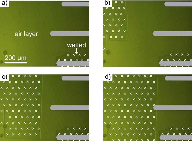
Figure 4.
Photographic images of a submerged air-retaining sample with micro-pillars taken over a duration of 15 minutes using the AFM camera. A chip holding three cantilevers (schematically indicated in grey) was installed, as can be seen on the right portion of each image. White crosses indicate areas that have been wetted. a) In the beginning, the surface shows air retention, with the exception of the lower right area. b) After about 3 minutes, a sudden change of the wetting state occurred, as the air layer collapsed on the upper left. c, d) The collapse occurred stepwise and erratically, propagating towards the cantilevers. In all cases, the interfaces separating the wetted areas from the air-retaining areas followed exactly the alignment of the micro-pillars.

