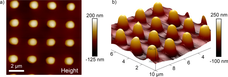
Figure 8.
a) AFM image in contact mode taken on a submerged air-retaining sample with an applied force of 6.4 nN. An area of 10 × 10 µm covering 16 pillars (bright spots) was scanned. b) The darker area between the pillars in a) indicates the shape of the air–water interface and can be better seen in the 3D representation of the data. Note that the x–y plane is scaled in micrometers and the height is scaled in nanometers.

