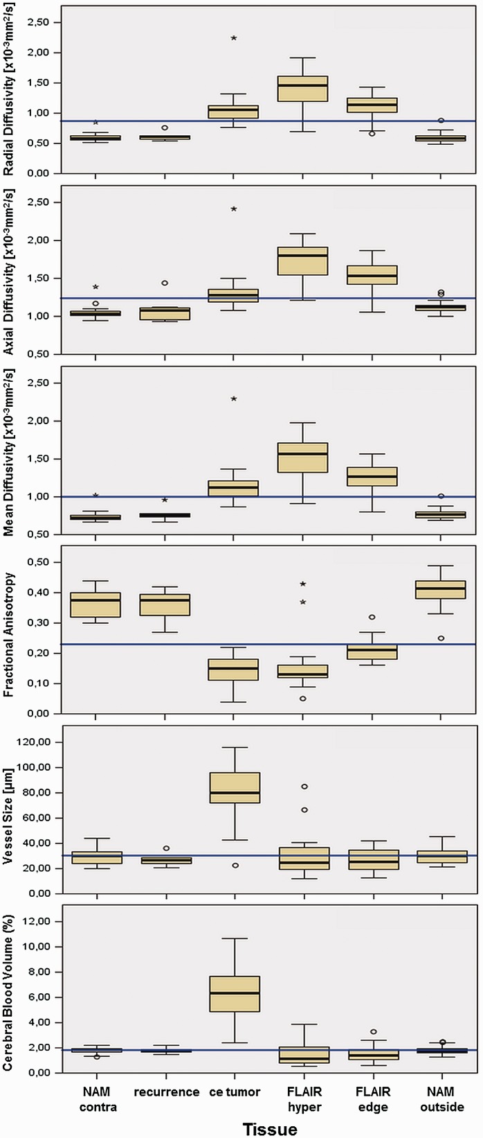
Figure 3.
Mesoscopic measures over investigated tissues.
Displayed from top to bottom are the median ± 2 SD of radial, axial and mean diffusivity (×10–3 mm2/s), fractional anisotropy (dimensionless), vessel size (µm), and CBV (%).
Displayed from left to right are the different investigated ROIs: contralateral normal-appearing matter (NAM_contra), recurrence, contrast-enhancing tumor (ce_tumour), T2/FLAIR hyperintense non-contrast-enhancing tumor (flair_hyper), non-contrast-enhancing tumor adjacent to the contrast enhancement (flair_edge), and normal-appearing matter adjacent but outside the tumor edge (NAM_outside). The total median of each mesoscopic measure is indicated in blue.
CBV: cerebral blood volume; FLAIR: fluid-attenuated inversion recovery.

