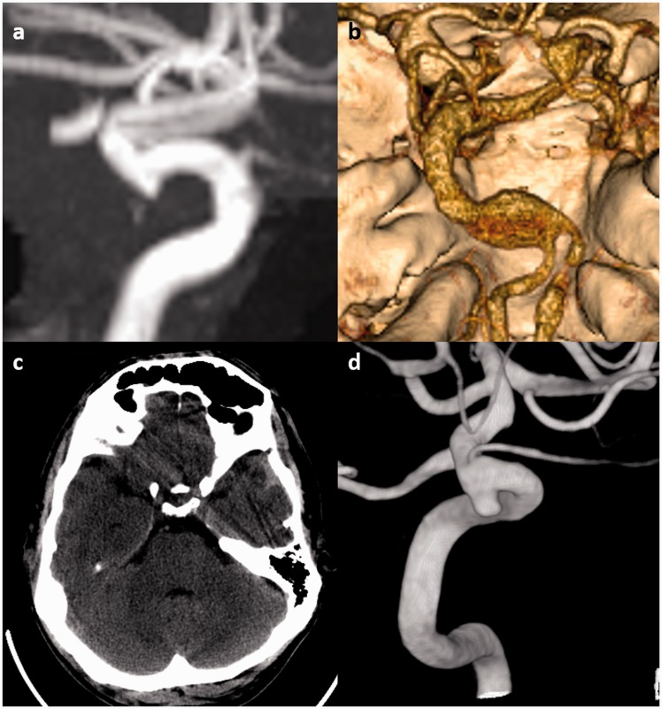
Figure 1.
Examples of intracranial aneurysms in connective-tissue diseases. (a) Incidental 3 mm supraclinoid internal carotid artery (ICA) aneurysm in a 64-year-old male Marfan syndrome patient. The aneurysm was managed conservatively. (b) An 8 mm dolichoectatic and fusiform aneurysm of the basilar artery in a 46-year-old female with Ehlers–Danlos syndrome. This aneurysm was managed conservatively because of the lack of good treatment options for this type of aneurysm in the setting of connective-tissue disease. (c) Non-contrast head computed tomography (CT) of a 50-year-old male with neurofibromatosis type 1 (NF1) shows a small amount of subarachnoid and subdural hemorrhage adjacent to the left temporal lobe. (d) Cerebral angiography demonstrates a 3 mm ruptured superior hypophyseal aneurysm that was effectively treated with stent-assisted coiling with no complications.

