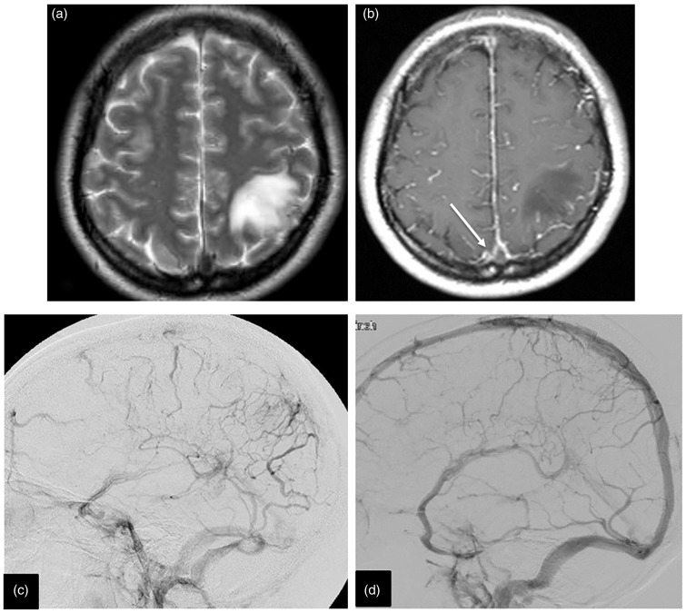
Figure 1.
(a) The lesion demonstrated by hyper intensity in T2-weighted MRI. (b) Contrast-enhanced magnetic resonance imaging showing an ‘empty delta sign’ in the superior sagittal sinus. (c) Angiography demonstrating SSS occlusion and left parietal venous congestion. (d) Three months after endovascular treatment, the angiography showed a good result of the recanalization.

