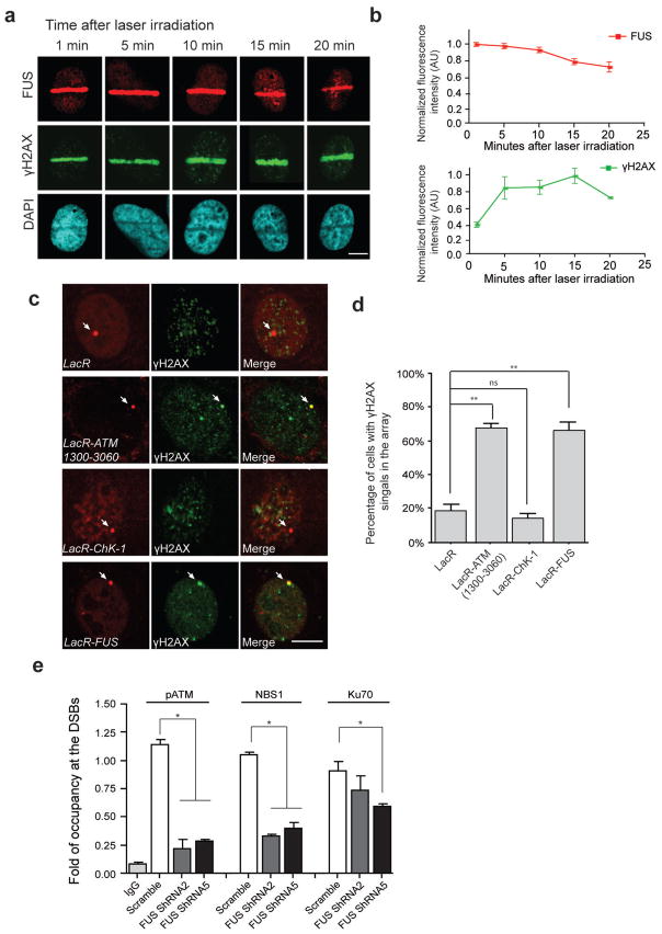
Figure 2. FUS is rapidly recruited to DSBs and is one of the earliest proteins to respond to DNA damage.
(a, b) Immunolabeling for endogenous FUS and γH2AX at the indicated time following the induction of DSBs via laser micro-irradiation of U2OS cells. FUS and γH2AX immunoreactive intensities within the irradiated area were normalized to the signal from the whole nucleus. Signals from 5–10 cells at each time point were averaged and normalized to the highest intensity across the time period. Scale bar: 8μm. (c, d) Immunofluorescent images were taken of NIH2/4 cells transfected with FUS or the indicated repair factors fused to LacR-mCherry (Red) or LacR- mCherry vector only. DNA damage response (DDR) activation is indicated by the presence of γH2AX foci (green). Arrow pointed mCherry signal indicates the tethering of the mCherry-fused proteins to the genomic loci of LacO array. Scale bar: 8 μm. The percentage of cells with colocalized γH2AX and LacR-mCherry signals in total LacR-mCherry expressing cells were calculated (mean ± SEM, n = 60–70, **P<0.01, unpaired t-test). (e) The occupancy of pATM, NBS1, and Ku70 at DSB sites was assessed using chromatin immunoprecipitation (ChIP) assays in U2OS-GFP cells following FUS shRNA-mediated knock-down (mean ± SEM, *P<0.05, one-way ANOVA).

