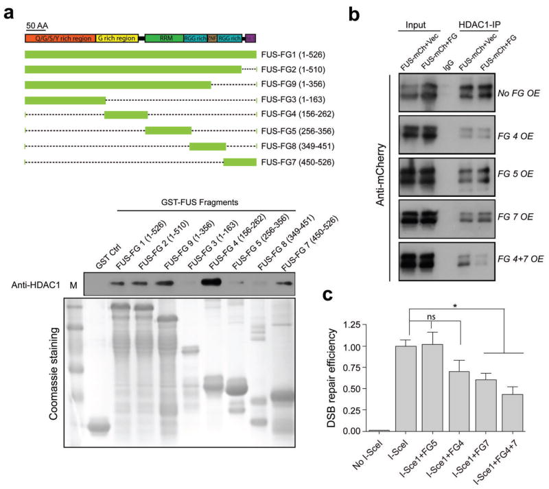
Figure 4. The G-rich and C-terminal domains of FUS directly interact with HDAC1, and this interaction is important for successful DSB repair.
(a) Top, schematic of various GST-FUS fragments (FG) that were generated corresponding to functional domains of FUS. RRM: RNA recognition motif; ZNF: zinc finger domain; RGG: the arginine, glycine rich domain; NLS: nuclear localization signal. Bottom, in vitro GST-pull down assay for mapping the functional domains of FUS that directly interact with HDAC1. GST tagged FUS fragments were incubated with recombinant HDAC1, pulled down using GST beads and blotted with anti-HDAC1 antibody. Protein inputs were shown by Coomassie blue staining of a duplicate gel. M: protein marker. (b) FUS fragments 4, 5, and 7 were co-transfected with full length FUS fused with mCherry (FUS-mCherry) in 293T cell as indicated and processed for immunoprecipitation with anti-HDAC1 antibody, and then blotted with anti-mCherry antibody. OE: overexpression. Vec: vector. Full-length blots are presented in Supplemental Fig. 10. (c) FUS fragments 4, 5, and 7 were over-expressed, together with I-SceI in U2OS-GFP cell line to evaluate DSB repair efficiency. GFP+ cells were analyzed by FACS. Repair efficiency was normalized to cells transfected with I-SceI alone (mean ± SEM, *P<0.05. unpaired t-test).

