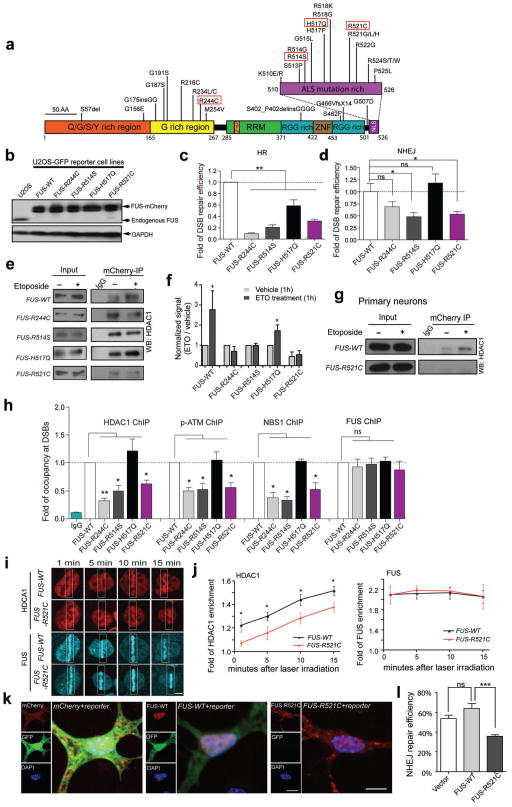
Figure 5. Cells carrying human fALS FUS mutations exhibit impaired DNA repair efficiency and a diminished FUS/HDAC1 interaction.
(a) FUS mutations identified in fALS patients. Red rectangles indicate those investigated in this study. (b) Anti-FUS blot of stable U2OS-GFP cell lines created by replacing endogenous FUS with mCherry tagged wild type or mutated FUS. (c, d) DNA repair assays of U2OS-GFP FUS cell lines (mean ± SEM, *P<0.05, **P< 0.01, unpaired t-test). (e, f) The interaction between FUS and HDAC1 in U2OS FUS cell lines. The signal intensity of etoposide-treated samples was compared to that of vehicle treated samples (mean ± SEM, *P<0.05, unpaired t-test). (g) The interaction between HDAC1 and FUS-WT or FUS-R521C in primary neurons. Full-length blots are presented in Supplemental Fig. 10. (h) ChIP-qPCR for analysis of the retention of HDAC1, pATM, NBS1 and FUS to the I-SceI created DSBs in U2OS-GFP FUS cell lines (mean ± SEM, *P< 0.05, **P< 01, unpaired t-test). (i, j) Representative images of endogenous HDAC1 (stained with anti-HDAC1 antibody) and overexpressed FUS-mCherry at the indicated time point following laser irradiation of FUS-WT or FUS-R521C U2OS cells. White boxes, lesioned area. Scale bar: 4 μm. Fluorescence intensities within lesioned area were normalized to those of the whole nucleus (mean ± SEM, n = 6–10, *P<0.05, unpaired test.). (k, l) DNA repair efficiency mediated by NHEJ was measured by the percentage of GFP+ cells in mCherry expressing cells (mean ± SEM, n = 60–80, ***P<0.001, unpaired t-test). Scale bar: 10 μm.

