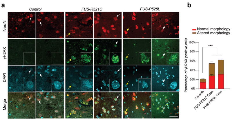
Figure 6. fALS patients harboring FUS mutations exhibit increased DNA damage.
(a, b) Representative images of γH2AX staining in human postmortem motor cortex sections of controls and ALS patients harboring FUS-R521C or FUS-P525L mutation. Higher magnificent image of arrow pointed cells was shown in the γH2AX and DAPI panels. Note that 48% and 54% of the NeuN+, γH2AX+ cells (white arrow) are morphologically normal and indistinguishable from surrounding NeuN+, γH2AX− cells in FUS-R521C and FUS-P525L brain sections, respectively. While the remaining cells exhibit altered morphology (yellow arrow). Error bar represents SD between slides. 5–6 images per slide, 5 slides each patient (mean ± SEM, ***P<0.001, one-way ANOVA). Scale bar: 20 μm.

