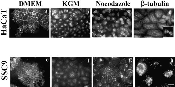
Figure 6.
Nocodazole effects on keratinocyte cell lines. HaCaT and SCC-9 cell lines cultured in DMEM containing 10% fetal bovine serum were transferred into KGM media to dissociate cells and then analyzed for the nocodazole effects. Cells were grown in DMEM (a and e), in KGM (b and c), and in KGM containing nocodazole (c and g) for 1 h and visualized by IF staining with anti-E-cadherin antibody. Nocodazole-induced cell-cell adhesions only in SCC-9 cells. β-Tubulin IF staining of HaCaT cells in KGM (d) and SCC9 cells in KGM containing nocodazole (h). Mitotic cells were frequently found in cells grown in KGM (d, arrow). Bar, 20 μm.

