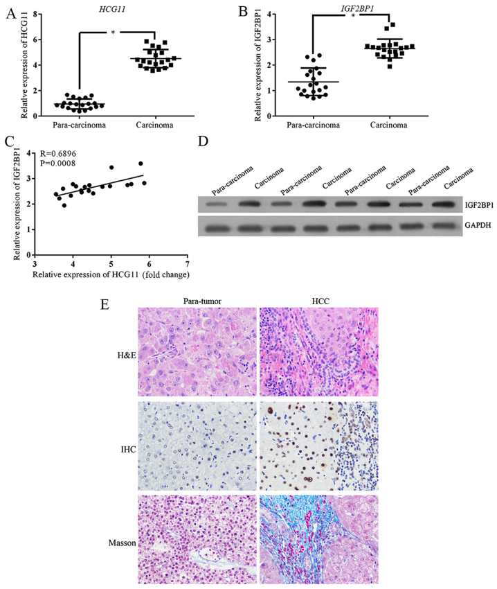
Figure 1.
The expression of HCG11 and IGF2BP1 is upregulated in HCC samples. (A) RT2-PCR validation shows upregulated expression of HCG11 in carcinoma tissues. (B) RT2-PCR validation shows upregulated expression of IGF2BP1 in carcinoma tissues. (C) A significantly positive association was detected between expression levels of HCG11 and IGF2BP1. (D) Upregulation of IGF2BP1 at protein level was detected between carcinoma and para-carcinoma tissues. (E) Histological staining illustration of HCC samples: following H&E staining, more infiltration cells could be observed in carcinoma tissues. IGF2BP1 expression were confirmed by IHC analysis. Additionally, higher fibrosis was also observed in carcinoma tissue by Masson. *P<0.05 vs. para-carcinoma tissues. Magnification, ×40.

