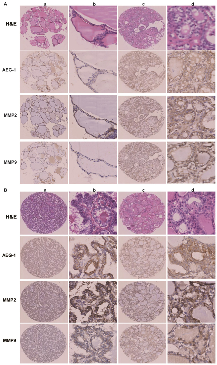
Figure 4.
AEG-1 expression positively correlated with MMP2 and MMP9 expression by immunohistochemistry. (A) AEG-1, MMP2 and MMP9 expressed lower level in normal thyroid tissue (a and b) than that in primary PTC without lymph node metastasis (c and d); a and c, ×40; b and d, ×200. (B) Both PTC with lymph node metastasis (a and b) and lymph node metastatic PTC (c and d) showed strong expression of AEG-1, MMP2 and MMP9, a and c, ×40; b and d, ×200.

