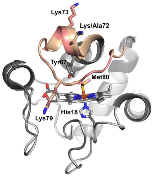
Figure 1.
Overlay of the structures of K72A Hu Cytc (PDB ID: 5TY3) and WT Hu Cytc (PDB ID: 3ZCF).25 The K72A variant is shown in light gray and WT Hu Cytc is shown in dark gray. The heme-crevice loop (Ω-loop D, residues 70–85) is highlighted in salmon for the K72A variant and tan for WT Hu Cytc. The heme and its environment, Met80, His18, and Tyr 67, are shown as stick models. The three lysine residues in Ω-loop D (Lys/Ala72, Lys73 and Lys79) are also shown as stick models.

