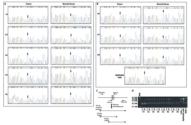
Figure 3. CAFs and normal fibroblasts from a panel of breast cancer biopsies are all heterozygous for the P16L polymorphism.
Direct amplicon sequencing of the GPER1 portion encoding the N-terminus of GPER of CAFs and normal fibroblasts from patients #1-5 with invasive carcinoma A. and from patients #6-8 with in-situ carcinoma and independent normal epithelial cells for comparison B. Note that all fibroblasts in this set of samples display a double peak with both C and T at the relevant position (arrows). C. Scheme of the analytical PCR protocol to confirm the presence of the C/T polymorphism indicated by sequencing. Using two common outer primers (Outer Fw and Outer Rv) and two allele-specific inner primers (Inner-C and Inner-T), PCR products of specific diagnostic sizes are generated. D. Image of an agarose gel for the genotyping of all samples with the PCR scheme of panel C. T and N stand for tumor (CAFs) and normal fibroblasts, respectively. Note that fibroblasts of patients #1-8 are heterozygous, CAFs_I homozygous for the variant with the T allele, and eptihelial cells from an unrelated breast carcinoma sample and SkBr3 cells are homozygous for the C allele.

