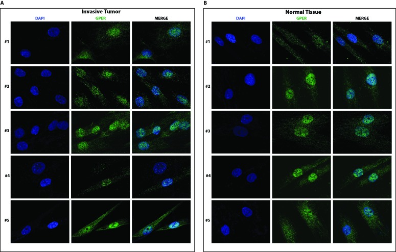
Figure 4. GPER localizes to both in the cytoplasm and the nucleus of CAFs A. and normal fibroblasts B. from invasive breast cancer biopsies.
Samples are from patients #1-5 with invasive breast carcinoma. Samples were stained with an anti-GPER antibody (green) and DAPI (blue), and analyzed by confocal microscopy. Immunofluorescence micrographs are representative of 10 different random fields.

