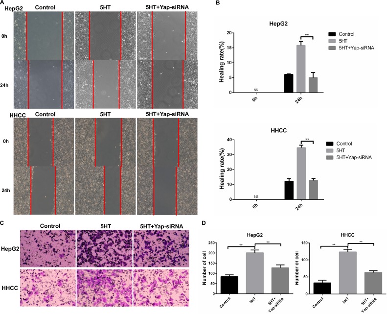
Figure 3. Yap promoted invasion and metastasis of HepG2 and HHCC cells.
(A–B) Inhibition of Yap induced slower wound closure in HepG2 and HHCC cells (A), and the healing rate was used for quantification (B). (C–D) Inhibition of Yap induced less cell penetration (C), and the penetrated cells were counted for quantification (D). ns: no statistical significance, **P < 0.01.

