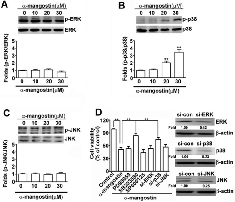
Figure 3. Effects of α-mangostin on MAPK pathways in cervical cancer cells.
HeLa cells were treated with increased concentrations of α-mangostin (0, 10, 20 and 30 μM) for 24 h. The levels of unphosphorylated and phosphorylated MAPK members, (A) ERK, (B) p38, and (C) JNK, were determined by immunoblotting. Quantitative results are shown in the bottom plot. (D) HeLa cells were pretreated with or without 50 μM MAPK inhibitors, PD98059 to ERK, SB203580 to p38, or SP600125 to JNK, for 2 h, and then treated with or without 20 μM α-mangostin for 24 h. Alternatively, HeLa cells were transfected with specific siRNAs against ERK, p38, or JNK for 24 h, and then the transfected cells were treated with 20 μM α-mangostin for 24 h. Cell viability was determined by MTT assay. **P < 0.01.

