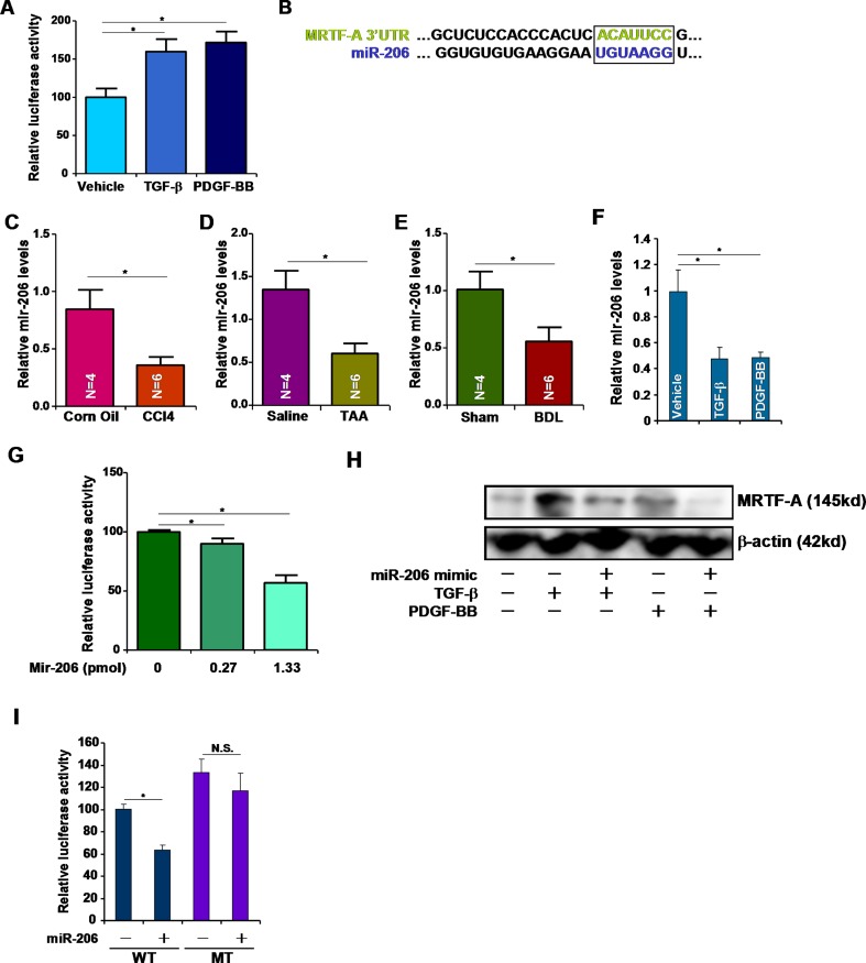
Figure 2. miR-206 regulates MRTF-A expression.
(A) A luciferase construct harboring the 3′-UTR of MRTF-A was transfected into HSC-T6 cells followed by with TGF-β or PDGF-BB for 24 hours. Luciferase activities were normalized by both protein concentration and GFP fluorescence. (B) Alignment of the 3′-UTR of the MRTF-A gene and miR-206. Matching sequences are highlighted and boxed. (C–E) Hepatic expression of miR-206 was examined by qPCR in (C) CCl4-, (D) TAA-, and (E) BDL-induced liver fibrosis in mice. (F) HSC-T6 cells were treated with TGF-β or PDGF-BB for 24 hours. Expression of miR-206 was examined by qPCR. (G) A luciferase construct harboring the 3′-UTR of MRTF-A was transfected into HSC-T6 cells with increasing doses of miR-206 mimic. Luciferase activities were normalized by both protein concentration and GFP fluorescence. (H) A miR-206 mimic was transfected into HSC-T6 cells followed by treated with TGF-β or PDGF-BB. MRTF-A expression was examined by Western. (I) A wild type (WT) or mutated (MT) MRTF-A reporter construct was transfected into HSC-T6 cells with or without miR-206. Luciferase activities were normalized by both protein concentration and GFP fluorescence.

